Upper Thigh Muscles Ct Anatomy : Muscles of the thigh - anterior | Hip muscles anatomy, Anatomy - A muscle of the medial thigh that originates on the pubis.
Upper Thigh Muscles Ct Anatomy : Muscles of the thigh - anterior | Hip muscles anatomy, Anatomy - A muscle of the medial thigh that originates on the pubis.. It arises by tendinous fibers from the anterior superior iliac spine and the upper half of the notch below it. Superior ramus of the pubis insertion: Reviewed by mary rodts, dnp. Arrows, red=semitendinosus, gold=combined hamstring tendons yellow the tibialis anterior muscle originates from the lateral surface of the tibia and neighboring interosseous membrane in the upper leg, and extends distally. Anatomical structures of the lower limb (hip, thigh, knee, leg, ankle and foot) and specific regions (compartment of the lower limb) are visible on cross section of the leg :
The hamstring muscles in the back of the thigh, the quadriceps muscles in the front, and the muscle strains usually happen when a muscle is stretched beyond its limit, tearing the muscle fibers. The thigh is the area between the hip and the knee joint. The muscles that move the forearm are located along the humerus, which include the triceps brachii, biceps brachii, brachialis, and brachioradialis. Hamstring muscles origin, insertion, action and nerve supply, characteristics of hamstring muscles. This injury frequently occurs near the point where the.
Presentation1.pptx, radiological anatomy of the thigh and leg. from image.slidesharecdn.com Anatomy of the human body. Muscles are named according to their shape, location, or a combination. The sparthos thigh compression sleeve provides compression as well as support for thigh muscles. Thigh muscle strains are common for people of all ages. There are different types of muscle, and some are controlled automatically by the autonomic nervous system. Anatomical structures of the lower limb (hip, thigh, knee, leg, ankle and foot) and specific regions (compartment of the lower limb) are visible on cross section of the leg : As the name implies they adduct the thigh at the hip joint. Thighs thigh muscles thigh actions and movements.
Superior ramus of the pubis insertion:
The iliopsoas is made up of two muscles that flex the thigh. The muscles of the thigh are arranged into three compartments. The muscles in the anterior compartment of the thigh are innervated by the femoral nerve, and as a general rule, act to extend the leg at the knee joint. Pain in the upper thigh can be difficult to diagnose because this area of the body contains many muscles, tendons, and ligaments. 2, vastus medialis & intermedius muscles. Thigh muscle strains are common for people of all ages. Want to learn more about it? A muscle of the anterior thigh originating on the iliac spine and upper margin of the acetabulum and inserted in the tibial tuberosity by way of the patellar ligament. The posterior compartment of the thigh contains the knee flexors and hip extensors.it has the following muscles, nerves and vessels: Create flashcards for free and quiz if you like muscles of upper limb, you might love these ideas. ·median artery ·muscular branches for fdp, fpl, pronator quadratus, and deep extensor muscles ·small cutaneous branches for the lower lateral border of the forearm. The sparthos thigh compression sleeve provides compression as well as support for thigh muscles. A muscle of the medial thigh that originates on the pubis.
In the upper back region, the trapezius, rhomboid major, and levator scapulae muscles anchor the scapula and clavicle to the spines of several vertebrae and in addition to moving the arm and pectoral girdle, muscles of the chest and upper back work together as a group to support the vital process of. In clinical anatomy the thigh muscles are divided into three groups: ·median artery ·muscular branches for fdp, fpl, pronator quadratus, and deep extensor muscles ·small cutaneous branches for the lower lateral border of the forearm. It inserts onto the linea aspera of the femur. Superior ramus of the pubis insertion:
Muscles of the hips and thighs | Human Anatomy and ... from s3-us-west-2.amazonaws.com Want to learn more about it? Reviewed by mary rodts, dnp. 2, vastus medialis & intermedius muscles. The gluteus medius muscle helps abducts the thigh along with the gluteus maximus, but can rotate the thigh inward where the gluteus maximus rotates the write down the muscles of the thigh in the table below and, for each, give the location of that muscle and what effect contracting that muscle has. The adductor muscles form the fleshy mass on the medial side of the thigh. In the upper back region, the trapezius, rhomboid major, and levator scapulae muscles anchor the scapula and clavicle to the spines of several vertebrae and in addition to moving the arm and pectoral girdle, muscles of the chest and upper back work together as a group to support the vital process of. It inserts onto the linea aspera of the femur. Lesser trochanter to linea aspera nerve supply:( double nerve.
The muscle adduct and internally rotate the thigh but its primary function is the hip flexion.
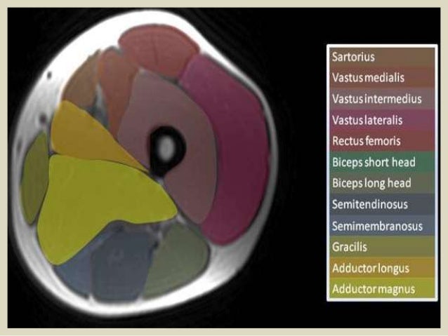
This bone is very thick and strong (due to the high proportion of bone tissue), and forms a ball and socket joint at the hip. Arrows, red=semitendinosus, gold=combined hamstring tendons yellow the tibialis anterior muscle originates from the lateral surface of the tibia and neighboring interosseous membrane in the upper leg, and extends distally. The pectineus muscle is a flat muscle that forms the base of the femoral triangle. The hamstring muscles in the back of the thigh, the quadriceps muscles in the front, and the muscle strains usually happen when a muscle is stretched beyond its limit, tearing the muscle fibers. This webpage presents the anatomical structures found on thigh mri. As the cursor is moved over a particular compartment of the lower. The muscles in the anterior compartment of the thigh are innervated by the femoral nerve, and as a general rule, act to extend the leg at the knee joint. Learn faster with these free muscle labeling diagrams. The muscles that move the forearm are located along the humerus, which include the triceps brachii, biceps brachii, brachialis, and brachioradialis. Muscles that move the shoulder and arm include the trapezius and serratus anterior. Thigh muscle strains are common for people of all ages. To better understand how to best target the arm musculature, let's first delve into basic anatomy. The muscles of the thigh are arranged into three compartments.
The muscles of the thigh are arranged into three compartments. It has a dual innervation, and thus can be considered a transitional. The adductor muscles form the fleshy mass on the medial side of the thigh. The pectineus muscle is a flat muscle that forms the base of the femoral triangle. Thighs thigh muscles thigh actions and movements.
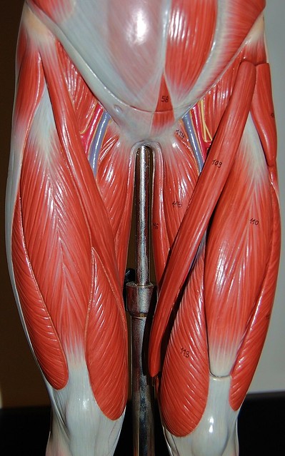
Muscles of the upper legs, anterior view | Flickr - Photo ... from c1.staticflickr.com Musculoskeletal anatomy, kinesiology, and palpation for manual therapists. There are different types of muscle, and some are controlled automatically by the autonomic nervous system. The muscle becomes stressed and tired after repeatedly doing the same motions over and over, leaving muscles fibers vulnerable to tears. 2, vastus medialis & intermedius muscles. The iliopsoas is made up of two muscles that flex the thigh. It inserts onto the linea aspera of the femur. The sparthos thigh compression sleeve provides compression as well as support for thigh muscles. The thigh is the area between the hip and the knee joint.
Muscular compartment, bones (tibia, fibula) and muscles.
Muscle anatomy of upper thigh, human muscles, muscle anatomy of upper thigh. Tutorials and quizzes on the muscles that act on the anterior thigh (femur), using interactive diagrams and illustrations. You've got an anterior compartment, medial, and posterior compartment and these are separated by the intermuscular this is this group of muscles here anteriorly in the thigh, obviously and these muscles are supplied by the femoral nerve. Case contributed by dr roberto schubert. Muscular compartment, bones (tibia, fibula) and muscles. Want to learn more about it? The thigh is the area between the hip and the knee joint. Muscles are groups of cells in the body that have the ability to contract and relax. As the name implies they adduct the thigh at the hip joint. Thigh muscle strains can occur when playing sports or participating in a daily activity. It inserts onto the linea aspera of the femur. As the cursor is moved over a particular compartment of the lower. It has a dual innervation, and thus can be considered a transitional.
Anatomy of the human body upper thigh anatomy. Pain in the upper thigh can be difficult to diagnose because this area of the body contains many muscles, tendons, and ligaments.
Share this post
1 Response to "Upper Thigh Muscles Ct Anatomy : Muscles of the thigh - anterior | Hip muscles anatomy, Anatomy - A muscle of the medial thigh that originates on the pubis."


This comment has been removed by the author.
ReplyDelete